THE E. COLI O157:H7 BACTERIA
A. Sources, Characteristics and Identification
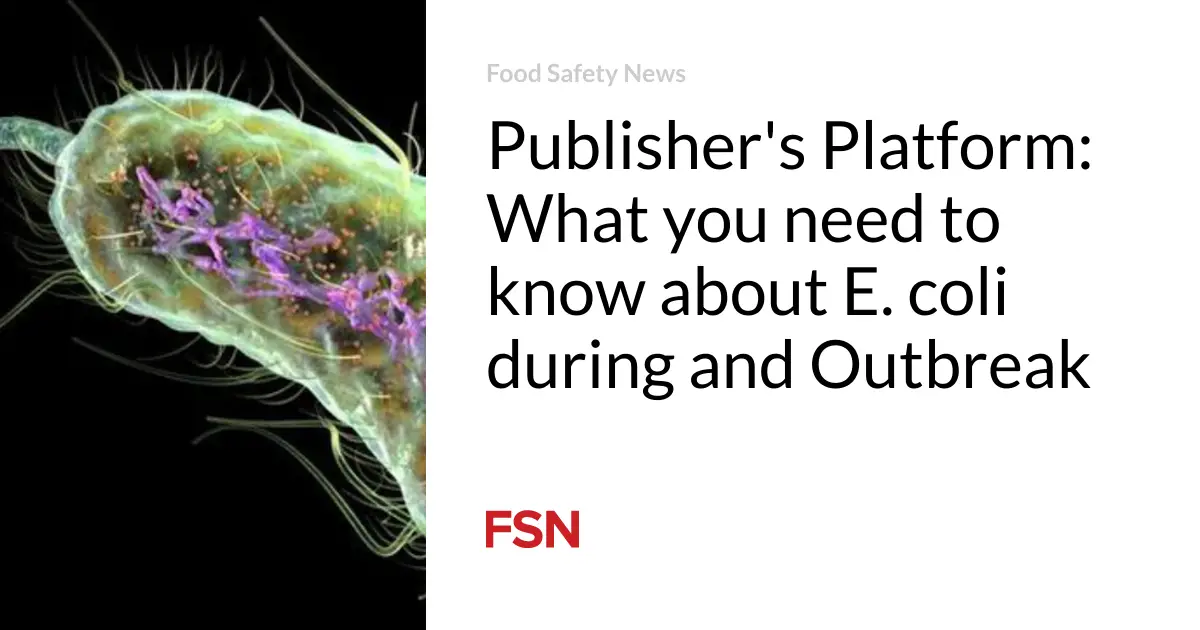
E. coli is an archetypal commensal bacterial species that lives in mammalian intestines. E. coli O157:H7 is one of thousands of serotypes Escherichia coli.[1] The combination of letters and numbers in the name of the E. coli O157:H7 refers to the specific antigens (proteins which provoke an antibody response) found on the body and tail or flagellum[2]respectively and distinguish it from other types of E. coli.[3] Most serotypes of E. coli are harmless and live as normal flora in the intestines of healthy humans and animals.[4] The E. coli bacterium is among the most extensively studied microorganism.[5] The testing done to distinguish E. coli O157:H7 from its other E. coli counterparts is called serotyping.[6] Pulsed-field gel electrophoresis (PFGE),[7] sometimes also referred to as genetic fingerprinting, is used to compare E. coli O157:H7 isolates to determine if the strains are distinguishable.[8] A technique called multilocus variable number of tandem repeats analysis (MLVA) is used to determine precise classification when it is difficult to differentiate between isolates with indistinguishable or very similar PFGE patterns.[9]
E. coli O157:H7 was first recognized as a pathogen in 1982 during an investigation into an outbreak of hemorrhagic colitis[10] associated with consumption of hamburgers from a fast food chain restaurant.[11] Retrospective examination of more than three thousand E. coli cultures obtained between 1973 and 1982 found only one (1) isolationwith serotype O157:H7, and that was a case in 1975.[12] In the ten (10) years that followed there were approximately thirty (30) outbreaks recorded in the United States.[13] This number is likely misleading, however, because E. coliO157:H7 infections did not become a reportable disease in any state until 1987 when Washington became the first state to mandate its reporting to public health authorities.[14] As a result, only the most geographically concentrated outbreak would have garnered enough notice to prompt further investigation.[15]
The E. coli O157:H7 Bacteria
E. coli O157:H7’s ability to induce injury in humans is a result of its ability to produce numerous virulence factors, most notably Shiga-like toxins.[16] Shiga toxin (Stx) has multiple variants (e.g. Stx1, Stx2, Stx2c), and acts like the plant toxin ricin by inhibiting protein synthesis in endothelial and other cells.[17] Shiga toxin is one of the most potent toxins known.[18] In addition to Shiga toxins, E. coli O157:H7 produces numerous other putative virulence factors including proteins, which aid in the attachment and colonization of the bacteria in the intestinal wall and which can lyse red blood cells and liberate iron to help support E. coli metabolism.[19]
E. coli O157:H7 evolved from enteropathogenic E. coli serotype O55:H7, a cause of non-bloody diarrhea, through the sequential acquisition of phage-encoded Stx2, a large virulence plasmid, and additional chromosomal mutations.[20]The rate of genetic mutation of E. coli O157:H7 indicates that the common ancestor of current E. coli O157:H7 clades[21] likely existed some 20,000 years ago.[22] E. coli O157:H7 is a relentlessly evolving organism,[23] constantly mutating and acquiring new characteristics, including virulence factors that make the emergence of more dangerous variants a constant threat.[24] The CDC has emphasized the prospect of emerging pathogens as a significant public health threat for some time.[25]
Although foods of a bovine origin are the most common cause of both outbreaks and sporadic cases of E. coliO157:H7 infections[26], outbreak of illnesses have been linked to a wide variety of food items. For example, produce has, since at least 1991, been the source of substantial numbers of outbreak-related E. coli O157:H7 infections.[27] Other unusual vehicles for E. coli O157:H7 outbreaks have included unpasteurized juices, yogurt, dried salami, mayonnaise, raw milk, game meats, sprouts, and raw cookie dough.[28]
According to a recent study, an estimated 93,094 illnesses are due to domestically acquired E. coli O157:H7 each year in the United States.[29] Estimates of foodborne acquired O157:H7 cases result in 2,138 hospitalizations and 20 deaths annually.[30] The colitis caused by E. coli O157:H7 is characterized by severe abdominal cramps, diarrhea that typically turns bloody within twenty-four (24) hours, and sometimes fevers.[31] The incubation period—which is to say the time from exposure to the onset of symptoms—in outbreaks is usually reported as three (3) to four (4) days, but may be as short as one (1) day or as long as ten (10) days.[32] Infection can occur in people of all ages but is most common in children.[33] The duration of an uncomplicated illness can range from one (1) to twelve (12) days.[34] In reported outbreaks, the rate of death is 0-2%, with rates running as high as 16-35% in outbreaks involving the elderly, like those have occurred at nursing homes.[35]
What makes E. coli O157:H7 remarkably dangerous is its very low infectious dose,[36] and how relatively difficult it is to kill these bacteria.[37] Unlike Salmonella, for example, which usually requires something approximating an “egregious food handling error, E. coli O157:H7 in ground beef that is only slightly undercooked can result in infection,”[38] as few as twenty (20) organisms may be sufficient to infect a person and, as a result, possibly kill them.[39] And unlike generic E. coli, the O157:H7 serotype multiplies at temperatures up to 44°F, survives freezing and thawing, is heat resistant, grows at temperatures up to 111°F, resists drying, and can survive exposure to acidic environments.[40]
And, finally, to make it even more of a threat, E. coli O157:H7 bacteria are easily transmitted by person-to-person contact.[41] There is also the serious risk of cross-contamination between raw meat and other food items intended to be eaten without cooking. Indeed, a principle and consistent criticism of the USDA E. coli O157:H7 policy is the fact that it has failed to focus on the risks of cross-contamination versus that posed by so-called improper cooking.[42] With this pathogen, there is ultimately no margin of error. It is for this precise reason that the USDA has repeatedly rejected calls from the meat industry to hold consumers primarily responsible for E. coli O157:H7 infections caused, in part, by mistakes in food handling or cooking.[43]
B. Hemolytic Uremic Syndrome (HUS)
E. coli O157:H7 infections can lead to a severe, life-threatening complication called hemolytic uremic syndrome (HUS).[44] HUS accounts for the majority of the acute deaths and chronic injuries caused by the bacteria.[45] HUS occurs in 2-7% of victims, primarily children, with onset five to ten days after diarrhea begins.[46] It is the most common cause of renal failure in children.[47] Approximately half of the children who suffer HUS require dialysis, and at least 5% of those who survive have long term renal impairment.[48] The same number suffers severe brain damage.[49] While somewhat rare, serious injury to the pancreas, resulting in death or the development of diabetes, can also occur.[50] There is no cure or effective treatment for HUS.[51]
HUS is believed to develop when the toxin from the bacteria, known as Shiga-like toxin (SLT), enters the circulation through the inflamed bowel wall.[52] SLT, and most likely other chemical mediators, attach to receptors on the inside surface of blood vessel cells (endothelial cells) and initiate a chemical cascade that results in the formation of tiny thrombi (blood clots) within these vessels.[53] Some organs seem more susceptible, perhaps due to the presence of increased numbers of receptors, and include the kidney, pancreas, and brain.[54] By definition, when fully expressed, HUS presents with the triad of hemolytic anemia (destruction of red blood cells), thrombocytopenia (low platelet count), and renal failure (loss of kidney function).[55]
As already noted, there is no known therapy to halt the progression of HUS. HUS is a frightening complication that even in the best American centers has a notable mortality rate.[56] Among survivors, at least five percent will suffer end stage renal disease (ESRD) with the resultant need for dialysis or transplantation.[57] But, “[b]ecause renal failure can progress slowly over decades, the eventual incidence of ESRD cannot yet be determined.”[58] Other long-term problems include the risk for hypertension, proteinuria (abnormal amounts of protein in the urine that can portend a decline in renal function), and reduced kidney filtration rate.[59] Since the longest available follow-up studies of HUS victims are 25 years, an accurate lifetime prognosis is not really available and remains controversial.[60] All that can be said for certain is that HUS causes permanent injury, including loss of kidney function, and it requires a lifetime of close medical-monitoring.
C. Other Medical Complications
Reactive Arthritis
The term reactive arthritis refers to an inflammation of one or more joints, following an infection localized at another site distant from the affected joints. The predominant site of the infection is the gastrointestinal tract. Several bacteria, including E. coli, induce septic arthritis.[61]The resulting joint pain and inflammation can resolve completely over time or permanent joint damage can occur.[62]
The reactive arthritis associated with Reiter Syndrome may develop after a person eats food that has been tainted with bacteria. In a small number of persons, the joint inflammation is accompanied by conjunctivitis (inflammation of the eyes), and urethritis (painful urination). Id. This triad of symptoms is called Reiter Syndrome.[63] Reiter Syndrome, a form of reactive arthritis, is an uncommon but debilitating syndrome caused by gastrointestinal or genitourinary infections. The most common gastrointestinal bacteria involved are Salmonella, Campylobacter, Yersinia, and Shigella. Reiter Syndrome is characterized by a triad of arthritis, conjunctivitis, and urethritis, although not all three symptoms occur in all affected individuals.[64]
Although the initial infection may not be recognized, reactive arthritis can still occur. Reactive arthritis typically involves inflammation of one joint (monoarthritis) or four or fewer joints (oligoarthritis), preferentially affecting those of the lower extremities; the pattern of joint involvement is usually asymmetric. Inflammation is common at entheses – i.e., the places where ligaments and tendons attach to bone, especially the knee and the ankle.
Salmonella has been the most frequently studied bacteria associated with reactive arthritis. Overall, studies have found rates of Salmonella-associated reactive arthritis to vary between 6 and 30%.[65] The frequency of postinfectious Reiter Syndrome, however, has not been well described. In a Washington State study, while 29% developed arthritis, only 3% developed the triad of symptoms associated with Reiter Syndrome.[66] In addition, individuals of Caucasian descent may be more likely those of Asian descent to develop reactive arthritis,[67] and children may be less susceptible than adults to reactive arthritis following infection with Salmonella.[68]
A clear association has been made between reactive arthritis and a genetic factor called the human leukocyte antigen (HLA) B27 genotype. HLA is the major histocompatibility complex in humans; these are proteins present on the surface of all body cells that contain a nucleus and are in especially high concentrations in white blood cells (leukocytes). It is thought that HLA-B27 may affect the elimination of the infecting bacteria or an individual’s immune response.[69]HLA-B27 has been shown to be a predisposing factor in one-half to over two-thirds of individuals with reactive arthritis.[70] While HLA-B27 does not appear to predispose to the initial infection itself, it increases the risk of developing arthritis that is more likely to be severe and prolonged. This risk may be slightly greater for Salmonella and Yersinia-associated arthritis than with Campylobacter, but more research is required to clarify this.[71]
Irritable Bowel Syndrome
A recently published study surveyed the extant scientific literature and noted that post-infectious irritable bowel syndrome (PI-IBS) is a common clinical phenomenon first-described over five decades ago.[72] The Walkerton Health Study further notes that:
Between 5% and 30% of patients who suffer an acute episode of infectious gastroenteritis develop chronic gastrointestinal symptoms despite clearance of the inciting pathogens.[73]
In terms of its own data, the “study confirm[ed] a strong and significant relationship between acute enteric infection and subsequent IBS symptoms.”[74] The WHS also identified risk-factors for subsequent IBS, including younger age; female sex; and four features of the acute enteric illness – diarrhea for > 7days, presence of blood in stools, abdominal cramps, and weight loss of at least ten pounds.[75]
Irritable bowel syndrome (IBS) is a chronic disorder characterized by alternating bouts of constipation and diarrhea, both of which are generally accompanied by abdominal cramping and pain.[76] In one recent study, over one-third of IBS sufferers had had IBS for more than ten years, with their symptoms remaining constant over time.[77] IBS sufferers typically experienced symptoms for an average of 8.1 days per month.[78]
As would be expected from a chronic disorder with symptoms of such persistence, IBS sufferers required more time off work, spent more days in bed, and more often cut down on usual activities, when compared with non-IBS sufferers.[79] And even when able to work, a significant majority (67%), felt less productive at work because of their symptoms.[80] IBS symptoms also have a significantly deleterious impact on social well-being and daily social activities, such as undertaking a long drive, going to a restaurant, or taking a vacation.[81] Finally, although a patient’s psychological state may influence the way in which he or she copes with illness and responds to treatment, there is no evidence that supports the theory that psychological disturbances in fact cause IBS or its symptoms.[82]
[1] E. coli bacteria were discovered in the human colon in 1885 by German bacteriologist Theodor Escherich. Feng, Peter, Stephen D. Weagant, Michael A. Grant, Enumeration of Escherichia coli and the Coliform Bacteria, in BACTERIOLOGICAL ANALYTICAL MANUAL (8th Ed. 2002), http://www.cfsan.fda.gov/~ebam/bam-4.html. Dr. Escherich also showed that certain strains of the bacteria were responsible for infant diarrhea and gastroenteritis, an important public health discovery. Id. Although the bacteria were initially called Bacterium coli, the name was later changed to Escherichia coli to honor its discoverer. Id.
[2] Not all E. coli are motile. For example, E. coli O157:H7 which lack flagella are thus E. coli O157:NM for non-motile.
[3] CDC, Escherichia coli O157:H7, General Information, Frequently Asked Questions: What is Escherichia coli O157:H7?, http://www.cdc.gov/ncidod/dbmd/diseaseinfo/escherichiacoli_g.htm.
[4] Marion Nestle, Safe Food: Bacteria, Biotechnology, and Bioterrorism, 40-41 (1st Pub. Ed. 2004).
[5] James M. Jay, MODERN FOOD MICROBIOLOGY at 21 (6th ed. 2000). (“This is clearly the most widely studied genus of all bacteria.”)
[6] Beth B. Bell, MD, MPH, et al. A Multistate Outbreak of Escherichia coli O157:H7-Associated Bloody Diarrhea and Hemolytic Uremic Syndrome from Hamburgers: The Washington Experience, 272 JAMA (No. 17) 1349, 1350 (Nov. 2, 1994) (describing the multiple step testing process used to confirm, during a 1993 outbreak, that the implicated bacteria were E. coli O157:H7).
[7] Jay, supra note 5, at 220-21 (describing in brief the PFGE testing process).
[8] Id. Through PFGE testing, isolates obtained from the stool cultures of probable outbreak cases can be compared to the genetic fingerprint of the outbreak strain, confirming that the person was in fact part of the outbreak. Bell, supra note 6, at 1351-52. Because PFGE testing soon proved to be such a powerful outbreak investigation tool, PulseNet, a national database of PFGE test results was created. Bala Swaminathan, et al. PulseNet: The Molecular Subtyping Network for Foodborne Bacterial Disease Surveillance, United States, 7 Emerging Infect. Dis. (No. 3) 382, 382-89 (May-June 2001) (recounting the history of PulseNet and its effectiveness in outbreak investigation).
[9] Konno T. et al. Application of a multilocus variable number of tandem repeats analysis to regional outbreak surveillance of Enterohemorrhagic Escherichia coli O157:H7 infections. Jpn J Infect Dis. 2011 Jan; 64(1): 63-5.
[10] “[A] type of gastroenteritis in which certain strains of the bacterium Escherichia coli (E. coli) infect the large intestine and produce a toxin that causes bloody diarrhea and other serious complications.” The Merck Manual of Medical Information, 2nd Home Ed. Online, http://www.merck.com/mmhe/sec09/ch122/ch122b.html.
[11] L. Riley, et al. Hemorrhagic Colitis Associated with a Rare Escherichia coli Serotype, 308 New. Eng. J. Med. 681, 684-85 (1983) (describing investigation of two outbreaks affecting at least 47 people in Oregon and Michigan both linked to apparently undercooked ground beef). Chinyu Su, MD & Lawrence J. Brandt, MD, Escherichia coli O157:H7 Infection in Humans, 123 Annals Intern. Med. (Issue 9), 698-707 (describing the epidemiology of the bacteria, including an account of its initial discovery).
[12] Riley, supra note 11 at 684. See also Patricia M. Griffin & Robert V. Tauxe, The Epidemiology of Infections Caused by Escherichia coliO157:H7, Other Enterohemorrhagic E. coli, and the Associated Hemolytic Uremic Syndrome, 13 Epidemiologic Reviews 60, 73 (1991).
[13] Peter Feng, Escherichia coli Serotype O157:H7: Novel Vehicles of Infection and Emergence of Phenotypic Variants, 1 Emerging Infect. Dis. (No. 2), 47, 47 (April-June 1995) (noting that, despite these earlier outbreaks, the bacteria did not receive any considerable attention until ten years later when an outbreak occurred 1993 that involved four deaths and over 700 persons infected).
[14] William E. Keene, et al. A Swimming-Associated Outbreak of Hemorrhagic Colitis Caused by Escherichia coli O157:H7 and Shigella Sonnei, 331 New Eng. J. Med. 579 (Sept. 1, 1994). See also Stephen M. Ostroff, MD, John M. Kobayashi, MD, MPH, and Jay H. Lewis, Infections with Escherichia coli O157:H7 in Washington State: The First Year of Statewide Disease Surveillance, 262 JAMA (No. 3) 355, 355 (July 21, 1989). (“It was anticipated the reporting requirement would stimulate practitioners and laboratories to screen for the organism.”)
[15] See Keene, supra note 14 at 583. (“With cases scattered over four counties, the outbreak would probably have gone unnoticed had the cases not been routinely reported to public health agencies and investigated by them.”) With improved surveillance, mandatory reporting in 48 states, and the broad recognition by public health officials that E. coli O157:H7 was an important and threatening pathogen, there were a total of 350 reported outbreaks from 1982-2002. Josef M. Rangel, et al. Epidemiology of Escherichia coli O157:H7 Outbreaks, United States, 1982-2002, 11 Emerging Infect. Dis. (No. 4) 603, 604 (April 2005).
[16] Griffin & Tauxe, supra note 12, at 61-62 (noting that the nomenclature came about because of the resemblance to toxins produced by Shigella dysenteries).
[17] Sanding K, Pathways followed by ricin and Shiga toxin into cells, Histochemistry and Cell Biology, vol. 117, no. 2:131-141 (2002). Endothelial cells line the interior surface of blood vessels. They are known to be extremely sensitive to E. coli O157:H7, which is cytotoxigenic to these cells making them a primary target during STEC infections.
[18] Johannes L, Shiga toxins—from cell biology to biomedical applications. Nat Rev Microbiol 8, 105-116 (February 2010). Suh JK, et al.Shiga Toxin Attacks Bacterial Ribosomes as Effectively as Eucaryotic Ribosomes, Biochemistry, 37 (26); 9394–9398 (1998).
[19] Welinder-Olsson C, Kaijser B. Enterohemorrhagic Escherichia coli (EHEC). Scand J. Infect Dis. 37(6-7): 405-16 (2005). See alsoUSDA Food Safety Research Information Office E. coli O157:H7 Technical Fact Sheet: Role of 60-Megadalton Plasmid (p0157) and Potential Virulence Factors, http://fsrio.nal.usda.gov/document_fsheet.php?product_id=225.
[20] Kaper JB and Karmali MA. The Continuing Evolution of a Bacterial Pathogen. PNAS vol. 105 no. 12 4535-4536 (March 2008). Wick LM, et al. Evolution of genomic content in the stepwise emergence of Escherichia coli O157:H7. J Bacteriol 187:1783–1791(2005).
[21] A group of biological taxa (as species) that includes all descendants of one common ancestor.
[22] Zhang W, et al. Probing genomic diversity and evolution of Escherichia coli O157 by single nucleotide polymorphisms. Genome Res 16:757–767 (2006).
[23] Robins-Browne RM. The relentless evolution of pathogenic Escherichia coli. Clin Infec Dis. 41:793–794 (2005).
[24] Manning SD, et al. Variation in virulence among clades of Escherichia coli O157:H7 associated with disease outbreaks. PNAS vol. 105 no. 12 4868-4873 (2008). (“These results support the hypothesis that the clade 8 lineage has recently acquired novel factors that contribute to enhanced virulence. Evolutionary changes in the clade 8 subpopulation could explain its emergence in several recent foodborne outbreaks; however, it is not clear why this virulent subpopulation is increasing in prevalence.”)
[25] Robert A. Tauxe, Emerging Foodborne Diseases: An Evolving Public Health Challenge, 3 Emerging Infect. Dis. (No. 4) 425, 427 (Oct.-Dec. 1997). (“After 15 years of research, we know a great deal about infections with E. coli O157:H7, but we still do not know how best to treat the infection, nor how the cattle (the principal source of infection for humans) themselves become infected.”)
[26] CDC, Multistate Outbreak of Escherichia coli O157:H7 Infections Associated with Eating Ground Beef—United States, June-July 2002, 51 MMWR 637, 638 (2002) reprinted in 288 JAMA (No. 6) 690 (Aug. 14, 2002).
[27] Rangel, supra note 15, at 605.
[28] Feng, supra note 13, at 49. See also USDA Bad Bug Book, Escherichia coli O157:H7, http://www.fda.gov/food/foodsafety/foodborneillness/foodborneillnessfoodbornepathogensnaturaltoxins/badbugbook/ucm071284.htm.
[29] Scallan E, et al. Foodborne illness acquired in the United States –major pathogens, Emerging Infect. Dis. Jan. (2011), http://www.cdc.gov/EID/content/17/1/7.htm.
[30] Id., Table 3.
[31] Griffin & Tauxe, supra note 12, at 63.
[32] Centers for Disease Control, Division of Foodborne, Bacterial and Mycotic Diseases, Escherichia coli general information, http://www.cdc.gov/nczved/dfbmd/disease_listing/stec_gi.html. See also PROCEDURES TO INVESTIGATE FOODBORNE ILLNESS, 107 (IAFP 5th Ed. 1999) (identifying incubation period for E. coli O157:H7 as “1 to 10 days, typically 2 to 5”).
[33] Su & Brandt, supra note 11 (“the young are most often affected”).
[34] Tauxe, supra note 25, at 1152.
[35] Id.
[36] Griffin & Tauxe, supra note 12, at 72. (“The general patterns of transmission in these outbreaks suggest that the infectious dose is low.”)
[37] V.K. Juneja, O.P. Snyder, A.C. Williams, and B.S. Marmer, Thermal Destruction of Escherichia coli O157:H7 in Hamburger, 60 J. Food Prot. (vol. 10). 1163-1166 (1997) (demonstrating that, if hamburger does not get to 130°F, there is no bacterial destruction, and at 140°F, there is only a 2-log reduction of E. coli present).
[38] Griffin & Tauxe, supra note 12, at 72 (noting that, as a result, “fewer bacteria are needed to cause illness that for outbreaks of salmonellosis”). Nestle, supra note 4, at 41. (“Foods containing E. coli O17:H7 must be at temperatures high enough to kill all of them.”) (italics in original)
[39] Patricia M. Griffin, et al. Large Outbreak of Escherichia coli O157:H7 Infections in the Western United States: The Big Picture, in RECENT ADVANCES IN VEROCYTOTOXIN-PRODUCING ESCHERICHIA COLI INFECTIONS, at 7 (M.A. Karmali & A. G. Goglio eds. 1994). (“The most probable number of E. coli O157:H7 was less than 20 organisms per gram.”) There is some inconsistency with regard to the reported infectious dose. Compare Chryssa V. Deliganis, Death by Apple Juice: The Problem of Foodborne Illness, the Regulatory Response, and Further Suggestions for Reform, 53 Food Drug L.J. 681, 683 (1998) (“as few as ten”) with Nestle, supra note 4, at 41 (“less than 50”). Regardless of these inconsistencies, everyone agrees that the infectious dose is, as Dr. Nestle has put it, “a miniscule number in bacterial terms.” Id.
[40] Nestle, supra note 4, at 41.
[41] Griffin & Tauxe, supra note 12, at 72. The apparent “ease of person-to-person transmission…is reminiscent of Shigella, an organism that can be transmitted by exposure to extremely few organisms.” Id. As a result, outbreaks in places like daycare centers have proven relatively common. Rangel, supra note 15, at 605-06 (finding that 80% of the 50 reported person-to-person outbreak from 1982-2002 occurred in daycare centers).
[42] See, e.g. National Academy of Science, Escherichia coli O157:H7 in Ground Beef: Review of a Draft Risk Assessment, Executive Summary, at 7 (noting that the lack of data concerning the impact of cross-contamination of E. coli O157:H7 during food preparation was a flaw in the Agency’s risk-assessment), http://www.nap.edu/books/0309086272/html/.
[43] Kriefall v. Excel, 265 Wis.2d 476, 506, 665 N.W.2d 417, 433 (2003). (“Given the realities of what it saw as consumers’ food-handling patterns, the [USDA] bored in on the only effective way to reduce or eliminate food-borne illness”—i.e., making sure that “the pathogen had not been present on the raw product in the first place.”) (citing Pathogen Reduction, 61 Fed. Reg. at 38966).
[44] Griffin & Tauxe, supra note 12, at 65-68. See also Josefa M. Rangel, et al. Epidemiology of Escherichia coli O157:H7 Outbreaks, United States, 1982-2002, 11 Emerging Infect. Dis. (No. 4) 603 (April 2005) (noting that HUS is characterized by the diagnostic triad of hemolytic anemia—destruction of red blood cells, thrombocytopenia—low platelet count, and renal injury—destruction of nephrons often leading to kidney failure).
[45] Richard L. Siegler, MD, The Hemolytic Uremic Syndrome, 42 Ped. Nephrology, 1505 (Dec. 1995) (noting that the diagnostic triad of hemolytic anemia, thrombocytopenia, and acute renal failure was first described in 1955). (“[HUS] is now recognized as the most frequent cause of acute renal failure in infants and young children.”) See also Beth P. Bell, MD, MPH, et al. Predictors of Hemolytic Uremic Syndrome in Children During a Large Outbreak of Escherichia coli O157:H7 Infections, 100 Pediatrics 1, 1 (July 1, 1997), at http://www.pediatrics.org/cgi/content/full/100/1/e12.
[46] Tauxe, supra note 25, at 1152. See also Nasia Safdar, MD, et al. Risk of Hemolytic Uremic Syndrome After Treatment of Escherichia coliO157:H7 Enteritis: A Meta-analysis, 288 JAMA (No. 8) 996, 996 (Aug. 28, 2002). (“E. coli serotype O157:H7 infection has been recognized as the most common cause of HUS in the United States, with 6% of patients developing HUS within 2 to 14 days of onset of diarrhea.”). Amit X. Garg, MD, MA, et al. Long-term Renal Prognosis of Diarrhea-Associated Hemolytic Uremic Syndrome: A Systematic Review, Meta-Analysis, and Meta-regression, 290 JAMA (No. 10) 1360, 1360 (Sept. 10, 2003). (“Ninety percent of childhood cases of HUS are…due to Shiga-toxin producing Escherichia coli.”)
[47] Su & Brandt, supra note 11.
[48] Safdar, supra note 46, at 996 (going on to conclude that administration of antibiotics to children with E. coli O157:H7 appeared to put them at higher risk for developing HUS).
[49] Richard L. Siegler, MD, Postdiarrheal Shiga Toxin-Mediated Hemolytic Uremic Syndrome, 290 JAMA (No. 10) 1379, 1379 (Sept. 10, 2003).
[50] Pierre Robitaille, et al., Pancreatic Injury in the Hemolytic Uremic Syndrome, 11 Pediatric Nephrology 631, 632 (1997) (“although mild pancreas involvement in the acute phase of HUS can be frequent”).
[51] Safdar, supra note 46, at 996. See also Siegler, supra note 49, at 1379. (“There are no treatments of proven value, and care during the acute phase of the illness, which is merely supportive, has not changed substantially during the past 30 years.”)
[52] Garg, supra note 46, at 1360.
[53] Id. Siegler, supra note 45, at 1509-11 (describing what Dr. Siegler refers to as the “pathogenic cascade” that results in the progression from colitis to HUS).
[54] Garg, supra note 46, at 1360. See also Su & Brandt, supra note 11, at 700.
[55] Garg, supra note46, at 1360. See also Su & Brandt, supra note 11, at 700.
[56] Siegler, supra note 45, at 1519 (noting that in a “20-year Utah-based population study, 5% dies, and an equal number of survivors were left with end-stage renal disease (ESRD) or chronic brain damage.”)
[57] Garg, supra note 46, at 1366-67.
[58] Siegler, supra note 45, at 1519.
[59] Id. at 1519-20. See also Garg, supra note 46, at 1366-67.
[60] Garg, supra note 46, at 1368.
[61] See J. Lindsey, “Chronic Sequellae of Foodborne Disease,” Emerging Infectious Diseases, Vol. 3, No. 4, Oct-Dec, 1997.
[62] Id.
[63] Id. See also Dworkin, et al., “Reactive Arthritis and Reiter’s Syndrome following an outbreak of gastroenteritis caused by Salmonella enteritidis,” Clin. Infect. Dis., 2001 Oct. 1;33(7): 1010-14; Barth, W. and Segal, K., “Reactive Arthritis (Reiter’s Syndrome),” American Family Physician, Aug. 1999, online at www.aafp.org/afp/990800ap/ 499.html.
[64] Hill Gaston JS, Lillicrap MS. (2003). Arthritis associated with enteric infection. Best Practices & Research Clinical Rheumatology. 17(2):219-39.
[65] Id.
[66] Dworkin MS, Shoemaker PC, Goldoft MJ, Kobayashi JM, “Reactive arthritis and Reiter’s syndrome following an outbreak of gastroenteritis caused by Salmonella enteritidis. Clin. Infect. Dis. 33(7):1010-14.
[67] McColl GJ, Diviney MB, Holdsworth RF, McNair PD, Carnie J, Hart W, McCluskey J, “HLA-B27 expression and reactive arthritis susceptibility in two patient cohorts infected with Salmonella Typhimurium,” Australian and New Zealand Journal of Medicine 30(1):28-32 (2001).
[68] Rudwaleit M, Richter S, Braun J, Sieper J, “Low incidence of reactive arthritis in children following a Salmonella outbreak,” Annals of the Rheumatic Diseases. 60(11):1055-57 (2001).
[69] Hill Gaston and Lillicrap, supra Note 7.
[70] Id.; Barth WF, Segal K., “Reactive arthritis (Reiter’s syndrome),” American Family Physician, 60(2):499-503, 507 (1999).
[71] Hill Gaston and Lillicrap, supra Note 7.
[72] J. Marshall, et al., Incidence and Epidemiology of Irritable Bowel Syndrome After a Large Waterborne Outbreak of Bacterial Dysentery, Gastro., 2006; 131; 445-50 (hereinafter “Walkerton Health Study” or “WHS”). The WHS followed one of the largest E. coli O157:H7 outbreaks in the history of North America. Contaminated drinking water caused over 2,300 people to be infected with E. coli O157:H7, resulting in 27 recognized cases of HUS, and 7 deaths. Id. at 445. The WHS followed 2,069 eligible study participants. Id. For Salmonella specific references, seeSmith, J.L., Bayles, D.O., Post-Infectious Irritable Bowel Syndrome: A Long-Term Consequence of Bacterial Gastroenteritis, Journal of Food Protection. 2007:70(7);1762-69.
[73] Id. at 445 (citing multiple sources).
[74] WHS, supra note 34, at 449.
[75] Id. at 447.
[76] A.P.S. Hungin, et al., Irritable Bowel Syndrome in the United States: Prevalence, Symptom Patterns and Impact, Aliment Pharmacol. Ther. 2005:21 (11); 1365-75.
[77] Id.at 1367.
[78] Id.
[79] Id. at 1368.
[80] Id.
[81] Id.
[82] Amy Foxx-Orenstein, DO, FACG, FACP, IBS—Review and What’s New, General Medicine 2006:8(3) (Medscape 2006) (collecting and citing studies). Indeed, PI-IBS has been found to be characterized by more diarrhea but less psychiatric illness with regard to its pathogenesis. SeeNicholas J. Talley, MD, PhD, Irritable Bowel Syndrome: From Epidemiology to Treatment, from American College of Gastroenterology 68th Annual Scientific Meeting and Postgraduate Course (Medscape 2003).


